On the median surface of the hemisphere, the major sulci separating lobes are the cingulate, parietooccipital, and also security (Figs. 16-4 and 16-5). One links the medial end of the main sulcus with the cingulate sulcus; the other joins the parietooccipital sulcus with the preoccipital notch. This mix of sulci and also lines separates the 4 wattles noted previously, plus the limbic wattle, on the median surface of the hemisphere (Fig. 16-4). On the basis of the setup of significant sulci, the cortex is separated right into 6 wattles, 5 of which are revealed externally of the analytical hemisphere and also one lies internal to the side sulcus. 4 of these lobes are named according to the overlying bones of the skull. It consists of 3 crucial segments, the cortex, the white matter, and also the subcortical structures.
- For telencephalon induction, this experiment would be technically challenging to carry out in the mouse, although perhaps possible in the chick.
- On day 1, wild kind fish were put in a white polymer storage tank, and also United States was given 10 s after CS got on.
- The human nerve system is separated right into the central nervous system and also the peripheral worried system.
- Identifying the forerunners at various beginning days additionally supplied information on the birthdates of the OT neurons, which were previously unknown for the computer mouse.
The amygdala (from the Greek word amygdale, indicating ‘almond’) is a small, almond-shaped cluster of nerve cells near one end of the hippocampus. Like the hippocampus, the amygdala contributes in memory and feeling, especially fear. People with damages to the amygdala are incapable to experience concern, also in circumstances where anxiety is appropriate. The human brain is made from over 100 billion afferent neuron that make trillions of links. Yet out of this complexity, scientists who examine the mind have actually been able to recognize distinctive structures, and they have even started to see how these frameworks are organized right into systems.
Division Of The Telencephalon Right Into Extra Hemisphere Sections
Monuki ES, Concierge FD, Walsh CA. Patterning of the dorsal telencephalon and also cortex by a roofing system plate-Lhx2 pathway. Hamasaki T, Leingartner A, Ringstedt T, O’Leary DD. EMX2 regulates dimensions as well as positioning of the primary sensory and also electric motor locations in neocortex by direct requirements of cortical progenitors. This paper gave the very first effort to describe exactly how expression slopes of 2 transcription variables, EMX2 and PAX6, develop area identity in the telencephalon.
These consist of the central sulcus between the parietal and also frontal wattles; the side sulcus in between the temporal, frontal, and also parietal lobes; as well as the parietooccipital sulcus in between the parietal and occipital lobes. The 6 major lobes consist of the frontal lobe, parietal wattle, temporal wattle, occipital wattle, insular lobe, as well as limbic wattle. The abovementioned surfaces are the medial surface, superolateral surface, and also a substandard surface. The equivalent boundaries are the premium margin and also inferolateral margin.
The Feature Of The Precentral Gyrus
The wattles of the cerebral cortex include the frontal, temporal, occipital, and parietal lobes. Figure 1.17 E is a section taken at the degree of the junction of the midbrain with the diencephalon. Notice that the plane of section varies from those of the previous sections.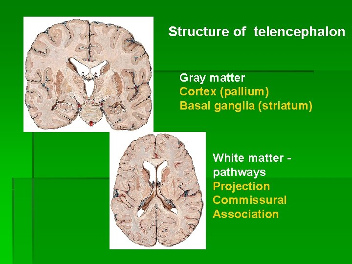
More details concerning any type of one of the major divisions above can be discovered by clicking the web link above or on the ideal area in the representation above. Crossley PH, Martin GR. The computer mouse Fgf8 gene inscribes a household of polypeptides and is shared in areas that guide outgrowth and also patterning in the establishing embryo. Although previous work by this group and also others had actually demonstrated a demand for LHX2 in the growth of the neocortex, this paper definitively demonstrated a total failing in neocortical specification in Lhx2 −/ − mice. The searching for suggests that LHX2 acts high in the hierarchy of neocortical induction, as well as supplies an entrance point for recognizing neocortical spec.
The Generation And Also Pattern Of Lge Forerunners Is Regular In The Absence Of Sox1
We followed the Sox1M1/ βgeo possible OT nerve cells with X-gal to establish whether they were capable of contributing to the OT. in HoHe embryos. ( F– M) 100-μm coronal sections were stained with X-gal to identify cells with Sox1 promoter activity. ( F– I) reveal Sox1βgeo/+ forebrain areas from E13 to birth revealing normal migration of Sox1-expressing cells from the VZ to the website of the OT, including striatal bridges. ( J– M) show sections of Sox1βgeo/ M1 forebrain, revealing lack of X-gal discoloration in the OT and also the striatal bridges. They appear as highly complicated masses of noodle that are arranged right into two rather balanced folded up frameworks.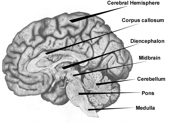
Really early in development, when the neural plate is curling right into the neural tube, mesodermic cells adapts a grid on each side of the developing tube as well as is separated into segments, which will at some point form the ribs as well as vertebrae. These sectors are referred to as somites, and also determine the development as well as gathering of neurons to develop the electric motor ganglions of the spine. The frontal eye field in people is located in the midsts of the precentral sulcus, in the cortex developing the rostral financial institution of the precentral sulcus, and prolonging onto the surface of the middle frontal gyrus (Fig. 16-9). This cortical location is mostly coextensive with Brodmann area 6 and also reaches the transitional area between areas 6 and 8 in the most back section of the middle frontal gyrus (Fig. 16-9). The present ideal proof in humans supports the view that the frontal eye field lies primarily in area 6.
Cortex
The telencephalon begins to arise in beginning growth at about 5 weeks. Currently, the nervous system contains tube-shaped item of tissue called the neural tube. The neural tube begins to establish swellings that will certainly later develop into vital structures in the nervous system. The swelling that kinds at the farthest end of the neural tube is called the telencephalon (telencephalon is Greek for “far brain”). The angular gyrus, which winds itself around the end of the premium temporal sulcus in an arc-like style, moderates in between the additional auditory cortex and second aesthetic cortex. This clarifies the value of the parietal association cortex with respect to features such as checking out or creating.
Layout showing the primary neighborhoods of the beginning animal mind. Drooping or peripheral paralysis is identified by a lack of tendon reflexes, whereas, on the other hand, the polysynaptic reflexes are not typically influenced. On top of that, flaccid paralysis leads to neurogenic degeneration of the muscles. The muscles are usually not influenced in abnormal paralysis as an outcome of the enhanced muscle tone.
Cortical Neuroanatomy
Along with the diencephalon, the telencephalon develops from the prosencephalon, the primitive forebrain 1. Short-term or working memory involves organization areas of the cortex, especially the dorsolateral prefrontal cortex, as well as the hippocampus. Abnormal paralysis manifests medically in regards to overstated proprioceptive reflexes as well as undermined or missing polysynaptic reflexes, as an example, along with other existing pathological reflexes. The Babinski sign is one example of a pathological reflex, expressed as dorsiflexion of the huge toe when the lateral plantar surface is scrubed.