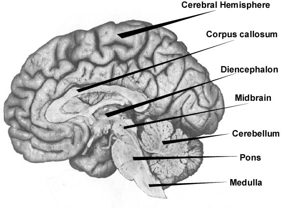
On the medial surface of the hemisphere, the major sulci dividing lobes are the cingulate, parietooccipital, and also security (Figs. 16-4 and also 16-5). One connects the median end of the main sulcus with the cingulate sulcus; the other joins the parietooccipital sulcus with the preoccipital notch. This combination of sulci and lines divides the 4 wattles kept in mind formerly, plus the limbic lobe, on the median surface area of the hemisphere (Fig. 16-4). On the basis of the setup of major sulci, the cortex is split into 6 lobes, five of which are subjected on the surface of the cerebral hemisphere and also one lies interior to the side sulcus. 4 of these wattles are named according to the superior bones of the skull. It includes three essential segments, the cortex, the white matter, and the subcortical frameworks.
- Not just the analytical sulci however likewise the mind convolutions increase the surface area of the mind.
- The two-way active evasion conditioning was likewise performed as blind experiments, in which the fish identities were not known to the experimenter (Fig. 6).
- These sectors are called rhombomeres and are classified r1 to r8 from anterior to caudal.
- The cerebrum is split by the medial longitudinal fissure into 2 cerebral hemispheres, the right and also the left.
- It also supplies spots for situating cores and also tracts associated with sensory and also electric motor functions.
The amygdala (from the Greek word amygdale, indicating ‘almond’) is a small, almond-shaped cluster of neurons near one end of the hippocampus. Like the hippocampus, the amygdala plays a role in memory and feeling, particularly fear. People with damages to the amygdala are not able to experience anxiety, even in circumstances where worry is appropriate. The human mind is made of over 100 billion afferent neuron that make trillions of links. Yet out of this intricacy, researchers that study the brain have been able to recognize distinct frameworks, and also they have actually even begun to see just how these structures are arranged into systems.
Developmental Neuroscience
Additionally, it regulates main sensory info crucial for discerning sensory experiences and the regulation of consciousness. The hypothalamus plays an important duty in the regulation of vegetative features, as it mediates in between mind functions, the internal secretion system, and the free nervous system. The so-called large mind includes the older part, the diencephalon, as well as the more youthful part, telencephalon. The thalamus and also hypothalamus lie in the location of the diencephalon. Bai CB, Stephen D, Joyner AL. All computer mouse forward spine patterning by hedgehog is Gli reliant and includes an activator function of Gli3. Furuta Y, Piston DW, Hogan BLM. Bone morphogenetic healthy proteins as regulators of dorsal forebrain advancement.
On day 5, the fish successfully escaped to one more compartment after CS got on. Herein, we recognized a subpopulation of nerve cells in the zebrafish Dm essential for anxiety conditioning. We suggest that these are practical matchings of nerve cells in the animal pallial amygdala, moderating the conditioned stimulation– unconditioned stimulus association. Thus, the research study develops a basis for comprehending the transformative conservation and also diversity of practical neural circuits moderating fear conditioning in animals. A cerebral hemisphere is made up of surface gray matter called cortex, underlying white issue and also deep masses of gray matter that are jointly called basic centers. Each side ventricle interacts with the third ventricle though an interventricular foramen.
Identity As Well As Migration Of Nerve Cells In The Vs
The wattles of the cerebral cortex consist of the frontal, temporal, occipital, and parietal wattles. Figure 1.17 E is a section taken at the level of the junction of the midbrain with the diencephalon. Notice that the plane of area varies from those of the previous areas.
In Shh −/ − mouse embryos, the telencephalon is minimized in size and also ventral cell kinds are lost17,23– 25. In double-mutant mice that lack both Shh and Gli3, however, ventral patterning is largely rescued16,26,27. Therefore, SHH restricts the dorsalizing function of GLI3 and also controls the positioning of the dorsoventral border. Therefore, SHH promotes forward identification by protecting against dorsalization of the telencephalon, instead of by straight promoting ventral cell personality.
Basic Cisterns Subarachnoid Tanks
For instance, the hands and the tongue possess a relatively huge region of innervation in the homunculus, whereas the upper legs, for example, just have a relatively small area. For example, an injury in the location of the parasagittal cortical area, which is provided by the anterior analytical artery, leads to a spastic paralysis of the legs. A long-term flaccid paralysis occurs when the 2nd motor nerve cell is damaged. These 2 kinds of paresis can be identified on the basis of scientific criteria. To this end, it receives afferents from the main and additional aesthetic cortex and supplies efferents to the mind muscular tissue nuclei of the cranial nerves III, IV and VI, by means of the remarkable colliculus.
It is bordered by a round crevice as well as is covered by parts of the surrounding wattles that construct the lid. The median temporal curvature is located listed below the upper curvature, as well as, in the back, it is extended by the angular curvature. The lower end of the fissure contains the frontal as well as frontoparietal sections of the operculum. In the aforementioned crevice of the dominant hemisphere, one can identify the Broca’s area that is in charge of speech. Fernandes M, Hébert JM. The ups and downs of holoprosencephaly, dorsal versus ventral patterning pressures.
Falces Of The Mind
As these computer mice reproduce consistently the Sox1-null phenotype with no evidence of a partial rescue, it is not likely that this is the result of incomplete expression of Sox1 from the Sox2 marketer in the forerunners. We found no difference in the degree and degree of expression of SOX1 protein in the VZ/SVZ of embryos with one copy of Sox1, whether it is shared from the Sox2 locus in HoHe or the endogenous Sox1 allele in Sox1βgeo/+ heterozygotes. Consequently, the introduction of VS/OT identification requires Sox1 expression in postmitotic cells. Our findings recommend that in other brain locations, subtype identification and movement might also be managed by the expression of transcription factors in postmitotic cells.
A much deeper understanding of how the fate of neural precursor cells is regulated in vivo will certainly offer new diagnostic and prognostic devices as well as healing targets for dealing with these problems. The Shh gene inscribes a secreted signalling healthy protein that belongs to the Drosophila healthy protein hedgehog. Shh is shared in the midline of the inceptive neural plate as well as remains to be expressed along the ventral midline of the CNS throughout development22 (FIG. 1).
Lateral view of human embryo at the beginning of the third and 5th week of gestation. An interesting, easily accessible and appealing book for any person who has even the least interest in exactly how the brain functions, but does not understand where to begin. Dingman weaves classic researches with modern research study into easily digestible sections, to offer a superb primer on the rapidly advancing area of neuroscience. Relying on the position of the animal it lies either in front or on top of the brainstem. In human beings, the cerebrum is the largest and also best-developed of the 5 major divisions of the brain.